Case Report | | Peer-Reviewed

Jugulocarotid Trauma in Civil Practice: About Three (3) Cases

Received: 20 May 2024    Accepted: 3 June 2024    Published: 19 June 2024
Views:       Downloads:
Abstract

Introduction: jugulocarotid trauma is rare and serious. They can be immediately life-threatening due to the impact of serious hemorrhage or obstruction of the upper airways. Objective: The objective of this work is to describe the clinical and therapeutic aspects of these lesions. Observations: The time taken to treat our patients was less than 12 hours in all our patients. A pressure dressing was placed in two patients. All our patients benefited from a preoperative assessment with Rh grouping and possibly a blood transfusion as needed. Exploratory cervicotomy was performed in all patients with, as appropriate, repair or ligation of the internal jugular vein and suture of the carotid wounds. All patients received antithrombotic treatment, antibiotic prophylaxis and analgesic treatment at the end of the procedure. The postoperative course was simple in all cases. Conclusion: vascular wounds of the neck constitute an emergency. Prehospital care is an essential step in the management of these lesions. Faced with massive hemorrhage, only immediate surgical exploration associated with resuscitation measures determines the survival of patients. In other cases, an imaging assessment may be necessary in stable patients.

Published in International Journal of Otorhinolaryngology (Volume 10, Issue 1)
DOI 10.11648/j.ijo.20241001.19
Page(s) 45-49
Creative Commons

This is an Open Access article, distributed under the terms of the Creative Commons Attribution 4.0 International License (http://creativecommons.org/licenses/by/4.0/), which permits unrestricted use, distribution and reproduction in any medium or format, provided the original work is properly cited.

Copyright

Copyright © The Author(s), 2024. Published by Science Publishing Group

Keywords

Jugulocarotid Trauma, Emergency, Exploratory Cervicotomy

1. Introduction
Penetrating cervical wounds are defined by the breakdown of the platysma muscle of the neck . The severity of ballistic trauma results, on the one hand, from the immediate and functional risks . Jugulocarotid lesions represent approximately 2 per 100,000 of all lesions during cervical trauma . Cervical projectile wounds are potentially serious injuries which are characterized by the difficulty of their initial management in the emergency setting and the specificity of the treatment methods for ballistic trauma . Zone II is the most frequently injured (47%) . Over the last ten years, advances in imaging (64-strip scanner) have radically changed the way these patients are managed, allowing non-invasive treatment for stable patients . Our clinical cases present particular mechanisms, namely the extremely rare occurrence of a firearm wound in the context of a domestic accident and an injury to the jugular vein in the context of AVP.
Our work aimed to review the clinical presentation of vascular neck wounds in three (3) cases and to report our attitude in the management of vascular lesions.
2. Observations
2.1. Observation 1
This was a 33-year-old female patient who presented with a cervical ballistic wound in the context of a domestic accident.
The onset of symptoms dates back 4 hours from her admission, marked by the sudden onset of an anterior cervical wound associated with massive hemorrhage without any notion of initial loss of consciousness when the patient lit the fire.
These symptoms prompted a consultation at the KABEZI hospital where blind clamping with hemostatic forceps was carried out, an isogroup-isorhesus blood transfusion with antitetanus serotherapy then the patient was transferred to Kamenge University Hospital for better care.
On cervical examination, we noted a circular right side-cervical wound presenting an entry orifice without exit orifice (figure 1). The neurological examination was normal.
The WHO activity index was rated at 0, we noted a slight conjunctivo-palmar pallor, BP: 90/60 mmHg, HR: 100 beats /min, FR: 20/min, T: 36.3° C, SaO2: 97%.
The frontal and lateral cervical x-ray revealed a foreign body of metallic density next to the C4-C5 vertebrae corresponding to the ball (figure 2), the cervical spine was intact. The CT could not be performed because it was not immediately available.
The exploratory cervicotomy carried out under general anesthesia allowed us to highlight a circular wound of approximately 1.5 mm located on the anterior edge of the sternocleidomastoid muscle at the level of its middle third. A Paul André "L" incision made along the edges of the wound allowed us to visualize and aspirate a large hematoma under the sternocleidomastoid. After aspiration and compression by compress pad; a complete section of the internal jugular vein has been note, whose lumen was blocked by a large clot and a linear breach of approximately 1cm on the right common carotid artery; the vagus nerve was intact.
Ligation of the internal jugular vein has been performed, after individualizing the vagus nerve, the carotid artery has been sutured with prolene 4.0 with a clamping and unclamping time of less than 10 min; the closure was carried out plan by plan.
Postoperatively, the patient was put on anticoagulant treatment, antibiotic prophylaxis and analgesic treatment.
The outcome was good and the patient was discharged after 10 days, without neurological complaints.
2.2. Observation 2
It was Mr. K, 57 years old, admitted to the ENT emergency room for post-traumatic neck injury.
His clinical history dates back two hours from his admission to the sudden occurrence of a neck injury following a road accident leading to massive hemorrhage without any notion of initial loss of consciousness.
The initial treatment provided at the scene of the accident consisted of a compressive dressing of the wound then the transfer of the patient to the Gabriel Touré University Hospital for treatment.
On cervical examination, we noted a right side-cervical wound covering zones I and II over approximately 10 cm with irregular edges and a large central hematoma.
The WHO activity index was rated at two, the conjunctiva colored. The pulse was 78 beats/min, BP was 110/70 mmHg.
The rest of the clinical examination was normal.
The hemoglobin level was 12 g/dl, hematocrit 36%, group O, rhesus positive.
Urgently an exploratory cervicotomy has been performed which allowed to note the section of the post edge of the sternocleidomastoid muscle and the anterior part of the scalene, after evacuation of the hematoma we noted a breach of the right jugular vein of approximately 2 cm in diameter at the height of half the muscle.
An initial compression of the wound has been performed, then a Paul André L incision, opening of the aponeurosis, dissection of the SCM, identification and clamping on both sides of the wound, we then evacuated the hematoma. After a lesion assessment, we dissected the IJV in the direction of the breach which measured approximately 2cm (figure 3). The breach has been sutured with prolene 4.0 (figure 4), after washing the wound, the closure was done in two layers on a drain.
At the end of the procedure, the patient received an iso-rhesus iso-group blood bag.
Postoperatively the patient was put on Amoxicillin: 50mg/Kg x2d, perfalgan (1g/6H), enoxaparin 0.4 (one subcutaneous injection per day), saline 0.9% (1 vial x2/d).
The postoperative course was simple. The control hemoglobin level was 13.6g/dl. Healing of the wound occurred around the 12th day.
No signs of thrombophlebitis, embolism or pyohemia have not been noted.
2.3. Observation 3
It involved a 38-year-old patient, a motorcycle taxi driver with no particular pathological history admitted to the department for cervical trauma following a deliberate stab wound.
The onset of symptoms dates back 2 hours from his admission, marked by the sudden onset of a latero-cervical neck wound associated with massive hemorrhage without any notion of initial loss of consciousness following a blow and intentional stab wound.
The initial treatment provided by the rescuers consisted of a compressive dressing of the wound then the transfer of the patient to the Gabriel Touré University Hospital for treatment.
On cervical examination we noted a right lateral-cervical wound of approximately 4 cm located in zone 2 of the neck, going from the posterior edge of the sternocleidomastoid muscle to the right lateral wing of the thyroid cartilage.
The WHO activity index was quoted as 0, BP: 95/60 mm Hg, HR: 100beat/min, FR: 2 1 cycles/min, T: 37.3°C.
The neurological examination was normal.
The exploratory cervicotomy carried out under general anesthesia allowed us to reveal a wound, approximately 4cm long, located in zone 2. A Paul André "L" incision made along the edges of the wound allowed us to visualize and to aspirate a large hematoma under the sternocleidomastoid. After aspiration and compression by compress pad; a linear wound of approximately 1.5 cm on the right carotid bifurcation hs been noted (figure 5).
After individualizing the vagus nerve, the carotid artery has been sutured with prolene 4.0 (figure 6) with a clamping and unclamping time of less than 10 min; the closure was carried out plan by plan.
Postoperatively, the patient was put on anticoagulant treatment, antibiotic prophylaxis and analgesic treatment.
The outcome was good and the patient was discharged after 10 days, without neurological complaints.
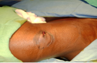
Figure 1. Projectile entry port.
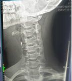
Figure 2. Cervical x-ray highlighted the projectile in the form of metallic hypodensity next to C4-C5.
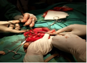
Figure 3. Dissection and clamping of the internal jugular vein.
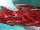
Figure 4. Reconstitution of the internal jugular vein.
Figure 5. Visualization of the carotid bifurcation gap.
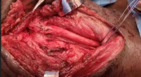
Figure 6. carotid suture with prolène 4.0.
3. Discussion
Jugulocarotid lesions represent approximately 1 to 2 per 100,000 of all neck injuries . In our department the incidence is estimated at approximately one case per year among all cervical trauma patients. This could be explained by the particular anatomical situation of these vessels; they are protected at the top by the bony mass of the base of the skull and at the bottom by the voluminous bundles of the sternocleidomastoid muscle.
The management of penetrating cervical wounds is classically based on the systematization of the neck into three anatomical zones according to the site of the injury . Zone I is bounded by the clavicle and the sternal fork below and by the lower border of the cricoid above. This area includes the brachiocephalic vessels, subclavian vessels, origin of the primary carotids, vertebral arteries, spinal cord, brachial plexus, trachea, esophagus, thoracic duct and pulmonary apices. Surgical exposure of this area is difficult due to the presence of the clavicle . Zone II is located between the lower edge of the cricoid and the mandibular angle. It contains the primary carotids which divide into internal and external carotids, the vertebral arteries, the internal and external jugular veins, the trachea, the esophagus, the spinal cord, the nerve roots. This area is easily accessible surgically . Zone III extends from the angle of the mandible to the base of the skull. It contains the internal carotids, the vertebral arteries, the pharynx, the spinal cord and its roots. This area is particularly difficult to expose surgically .
The systematization of the neck into three compartments (two lateral and one central compartment) seems relevant in the management of cervical vascular wounds . The lateral compartment is the site of vascular lesions; wounds in this compartment more often require surgical exploration. Central compartment damage is responsible for damage to the upper aerodigestive tract requiring initial orotracheal intubation, with initial monitoring in intensive care and possible delayed surgical management .
Prehospital management carried out by first aiders constitutes an essential step to ensure the survival of patients until the hospital environment . In our series, the initial treatment consisting of a compressive dressing of the wound and the release of the airway was very useful. The authors agree on the systematic surgical exploration of these lesions . Our clinical cases are an illustration of this. However, in hemodynamically stable patients, when conditions permit, angiography can be performed possibly coupled with embolization sessions Standard radiographs are useful when polyscreening with radiopaque munitions or when wearing fixed radiopaque dental equipment creating artifacts during the CT examination, but we cannot currently be satisfied with this sole assessment in a hospital center equipped with a CT scan . In our case, taking into account the insufficiency of the technical platform and the risks during travel, we carried out immediate surgical exploration. Arteriography is currently increasingly being replaced by CT angiography. It makes it possible to locate the projectile and, using an image subtraction technique, to objectify the vascular lesions without being hampered by metallic bodies. It diagnoses the type of vascular lesions: thromboses or spasms (reduction of vascular caliber, or even obstruction) and wounds or fistulas (leakage of the contrast product) .
The authors recommend paring with debridement, abundant washing of the wound with physiological saline and broad-spectrum antibiotic prophylaxis . The infection prevention protocol that we instituted was based on debridement, washing and antibiotic therapy.
4. Conclusion
Vascular wounds of the neck constitute an emergency. Prehospital care is an essential step in the management of these lesions. Faced with massive hemorrhage, only immediate surgical exploration associated with resuscitation measures determines the survival of patients. In other cases, an imaging assessment may be necessary in stable patients.
Abbreviations

C

Cervical Vertebra

Author Contributions
N’faly Konaté: Conceptualization, Data curation, Formal Analysis, Investigation, Methodology, Validation, Writing – original draft, Writing – review & editing
Sixte Nderagakura: Conceptualization, Data curation, Investigation, Writing – original draft
Kassim Diarra: Conceptualization, Data curation, Formal Analysis, Investigation, Methodology, Writing – original draft
Harouna Sanogo: Data curation, Investigation
Boubakary Guindo: Software
Kalifa Coulibaly: Formal Analysis, Investigation
Ibrahim Bouare: Data curation, Investigation
Mohamed Keïta: Conceptualization, Data curation, Investigatio, Software, Supervision, Validation
Conflicts of Interest
The authors declare no conflicts of interest.
References
[1] J. F. QUINOT, E. KAISER. Urgence devant une plaie cervicale [Emergency in front of a cervical wound]. EMC-Médecine 1. 2004, 157–164.
[2] KONÉ FI, KEITA MA, SOUMAHORO S, KONATÉ N, DIARRA K. Historical Case of Cervical Penetrating Wound: From First Aid to Surgical Intervention. Hindawi Case Reports in Otolaryngology. 2017. 3p.
[3] CESARE E, DRAOUA S, LEFORT H, TAZAROURTE K. Plaies pénétrantes du cou. [Penetrating areas of the neck.] SAMU de France urgence 2012.
[4] BALLIVET DE REGLOIX S, CRAMBERT A, MAURIN O, PONS Y, BONFORT G, CLEMENT P. Plaies cervicofaciales par projectiles [Head and neck games using projectiles]. EMC - Oto-rhino-laryngologie 2016; 11(2): 1-11 [Article 20-860-B-10].
[5] INABA K, MUNERA F, MCKENNEY, MG. The nonoperative management of penetrating internal jugular vein injury. J Vasc Surg 2006; 43: 77-80.
[6] Varghese A. Penetrating Neck Injury: A Case Report and Review of Management Indian J Surg 2013, 75(1): 43–46.
[7] ALTABAA K, FIORETTI E, VERILLAUD B. Prise en charge des plaies cervicales avec atteinte vasculaire. À propos de 30 cas [Management of cervical wounds with vascular involvement. About 30 cases]. In Annales Françaises d’ORL et de pathologie cervicofaciale. Paris 2013. 130(4): A53 https://doi.org/10.1016/J.aforl. 2013.06.138
[8] VAUDEY JG. Plaies et ligature de la veine jugulaire interne [Games and ligation of the internal jugular vein]. In Vaisseaux sanguins—Maladie. Paris 1890. 60 P.
[9] L. MARTIN. Traumatisme fermé des vaisseaux du cou [Trauma of closed vessels of the neck]. Prise en charge d’un polytraumatisé. Mapar 2002. 565-567.
[10] Mahmoodie M, Sanei B, Moazeni Bistgani M, Namgar M. Penetrating Neck Trauma: Review of 192 Cases. Arch Trauma Res. 2012 1(1): 4.
Cite This Article
  • APA Style

    Konaté, N., Nderagakura, S., Diarra, K., Sanogo, H., Guindo, B., et al. (2024). Jugulocarotid Trauma in Civil Practice: About Three (3) Cases. International Journal of Otorhinolaryngology, 10(1), 45-49. https://doi.org/10.11648/j.ijo.20241001.19

    Copy | Download

    ACS Style

    Konaté, N.; Nderagakura, S.; Diarra, K.; Sanogo, H.; Guindo, B., et al. Jugulocarotid Trauma in Civil Practice: About Three (3) Cases. Int. J. Otorhinolaryngol. 2024, 10(1), 45-49. doi: 10.11648/j.ijo.20241001.19

    Copy | Download

    AMA Style

    Konaté N, Nderagakura S, Diarra K, Sanogo H, Guindo B, et al. Jugulocarotid Trauma in Civil Practice: About Three (3) Cases. Int J Otorhinolaryngol. 2024;10(1):45-49. doi: 10.11648/j.ijo.20241001.19

    Copy | Download

  • @article{10.11648/j.ijo.20241001.19,
      author = {N’faly Konaté and Sixte Nderagakura and Kassim Diarra and Harouna Sanogo and Boubakary Guindo and Mohamed Ag El Ansari and Fatogoma Issa Kone and Naouma Cissé and Kalifa Coulibaly and Siaka Soumaoro and Kadiatou Sinkare and Ibrahima Bouare and Mohamed Keïta},
      title = {Jugulocarotid Trauma in Civil Practice: About Three (3) Cases
    },
      journal = {International Journal of Otorhinolaryngology},
      volume = {10},
      number = {1},
      pages = {45-49},
      doi = {10.11648/j.ijo.20241001.19},
      url = {https://doi.org/10.11648/j.ijo.20241001.19},
      eprint = {https://article.sciencepublishinggroup.com/pdf/10.11648.j.ijo.20241001.19},
      abstract = {Introduction: jugulocarotid trauma is rare and serious. They can be immediately life-threatening due to the impact of serious hemorrhage or obstruction of the upper airways. Objective: The objective of this work is to describe the clinical and therapeutic aspects of these lesions. Observations: The time taken to treat our patients was less than 12 hours in all our patients. A pressure dressing was placed in two patients. All our patients benefited from a preoperative assessment with Rh grouping and possibly a blood transfusion as needed. Exploratory cervicotomy was performed in all patients with, as appropriate, repair or ligation of the internal jugular vein and suture of the carotid wounds. All patients received antithrombotic treatment, antibiotic prophylaxis and analgesic treatment at the end of the procedure. The postoperative course was simple in all cases. Conclusion: vascular wounds of the neck constitute an emergency. Prehospital care is an essential step in the management of these lesions. Faced with massive hemorrhage, only immediate surgical exploration associated with resuscitation measures determines the survival of patients. In other cases, an imaging assessment may be necessary in stable patients.
    },
     year = {2024}
    }
    

    Copy | Download

  • TY  - JOUR
    T1  - Jugulocarotid Trauma in Civil Practice: About Three (3) Cases
    
    AU  - N’faly Konaté
    AU  - Sixte Nderagakura
    AU  - Kassim Diarra
    AU  - Harouna Sanogo
    AU  - Boubakary Guindo
    AU  - Mohamed Ag El Ansari
    AU  - Fatogoma Issa Kone
    AU  - Naouma Cissé
    AU  - Kalifa Coulibaly
    AU  - Siaka Soumaoro
    AU  - Kadiatou Sinkare
    AU  - Ibrahima Bouare
    AU  - Mohamed Keïta
    Y1  - 2024/06/19
    PY  - 2024
    N1  - https://doi.org/10.11648/j.ijo.20241001.19
    DO  - 10.11648/j.ijo.20241001.19
    T2  - International Journal of Otorhinolaryngology
    JF  - International Journal of Otorhinolaryngology
    JO  - International Journal of Otorhinolaryngology
    SP  - 45
    EP  - 49
    PB  - Science Publishing Group
    SN  - 2472-2413
    UR  - https://doi.org/10.11648/j.ijo.20241001.19
    AB  - Introduction: jugulocarotid trauma is rare and serious. They can be immediately life-threatening due to the impact of serious hemorrhage or obstruction of the upper airways. Objective: The objective of this work is to describe the clinical and therapeutic aspects of these lesions. Observations: The time taken to treat our patients was less than 12 hours in all our patients. A pressure dressing was placed in two patients. All our patients benefited from a preoperative assessment with Rh grouping and possibly a blood transfusion as needed. Exploratory cervicotomy was performed in all patients with, as appropriate, repair or ligation of the internal jugular vein and suture of the carotid wounds. All patients received antithrombotic treatment, antibiotic prophylaxis and analgesic treatment at the end of the procedure. The postoperative course was simple in all cases. Conclusion: vascular wounds of the neck constitute an emergency. Prehospital care is an essential step in the management of these lesions. Faced with massive hemorrhage, only immediate surgical exploration associated with resuscitation measures determines the survival of patients. In other cases, an imaging assessment may be necessary in stable patients.
    
    VL  - 10
    IS  - 1
    ER  - 

    Copy | Download

Author Information
  • ENT and Head and Neck Surgery Department, University Hospital Center “Gabriel Touré”, Bamako, Mali

  • ENT and Head and Neck Surgery Department, University Hospital Center “Kamenge”, Bujumbura, Burundi

  • ENT and Head and Neck Surgery Department, University Hospital Center “Gabriel Touré”, Bamako, Mali

  • ENT and Head and Neck Surgery Department, Reference Health Center, Kalaban Coro, Mali

  • ENT and Head and Neck Surgery Department, University Hospital Center “Gabriel Touré”, Bamako, Mali

  • ENT and Head and Neck Surgery Department, Commune VI Reference Health Center, Bamako, Mali

  • ENT and Head and Neck Surgery Department, University Hospital Center “Gabriel Touré”, Bamako, Mali

  • ENT and Head and Neck Surgery Department, University Hospital Center “Gabriel Touré”, Bamako, Mali

  • ENT and Head and Neck Surgery Department, University Hospital Center “Gabriel Touré”, Bamako, Mali

  • ENT and Head and Neck Surgery Department, University Hospital Center “Gabriel Touré”, Bamako, Mali

  • ENT and Head and Neck Surgery Department, University Hospital Center “Gabriel Touré”, Bamako, Mali

  • ENT and Head and Neck Surgery Department, University Hospital Center “Gabriel Touré”, Bamako, Mali

  • ENT and Head and Neck Surgery Department, Commune VI Reference Health Center, Bamako, Mali